Appendicular Skeleton Quiz
True / False
True / False
True / False
True / False
Appendicular Skeleton Labeling Errors That Break Sidedness and Articulation Logic
1) Mixing appendicular with axial bones
A frequent miss is labeling ribs, sternum, vertebrae, or skull bones on a limb diagram. Fix it by setting a boundary first: appendicular includes the girdles (clavicle, scapula, hip bones) plus the limb bones. If the structure is part of the thorax, spine, or skull, it is axial.
2) Calling the correct bone but the wrong side
Sidedness errors come from skipping anatomical position. Rebuild it mentally before reading landmarks: palms forward, thumbs lateral, feet forward. Then use direction cues instead of guesswork.
- Humerus: head is medial, olecranon fossa is posterior.
- Radius: broad distal end, styloid is lateral, aligns with the thumb.
- Ulna: trochlear notch faces anterior, olecranon is posterior.
- Tibia and fibula: tibial medial malleolus is medial, fibular lateral malleolus is lateral.
3) Naming a landmark without verifying the parent bone
Students often identify “acetabulum” or “glenoid cavity” correctly, then attach it to the wrong region. Use articulation logic: glenoid cavity pairs with humeral head, acetabulum pairs with femoral head, trochlea and capitulum are on the distal humerus.
4) Getting lost in carpals and tarsals
Memorized word chains fail on pictures. Start with regions and rows. Wrist bones sit in two rows, and the foot splits into hindfoot (talus, calcaneus), midfoot (navicular, cuboid, cuneiforms), and forefoot (metatarsals, phalanges).
Printable Appendicular Skeleton Quick Sheet: Girdles, Limb Bones, and High-Yield Landmarks
Print or save as PDF: Keep this sheet next to a lab model or diagram set so you can verify orientation and landmarks quickly while practicing.
What counts as appendicular
- Pectoral girdle: clavicle, scapula
- Upper limb: humerus, radius, ulna, carpals, metacarpals, phalanges
- Pelvic girdle: right and left hip bones (ilium, ischium, pubis fused)
- Lower limb: femur, patella, tibia, fibula, tarsals, metatarsals, phalanges
Fast sidedness rules (use anatomical position)
- Humerus: head points medial, olecranon fossa is posterior.
- Radius vs ulna: radius is lateral and has the larger distal end, ulna has the olecranon and trochlear notch.
- Tibia vs fibula: tibia is medial and weight-bearing, fibula is lateral and does not form the main knee joint surface.
- Femur: head points medial, linea aspera is posterior, patellar surface is anterior.
High-yield landmarks by bone
- Scapula: spine, acromion, coracoid process, glenoid cavity, inferior angle, medial border.
- Clavicle: sternal (medial) end, acromial (lateral) end.
- Humerus: greater and lesser tubercles, deltoid tuberosity, capitulum (lateral), trochlea (medial), medial and lateral epicondyles.
- Radius: head (proximal), radial tuberosity, ulnar notch (distal medial), styloid process (distal lateral).
- Ulna: olecranon, coronoid process, trochlear notch, radial notch (proximal lateral), styloid process (distal).
- Hip bone: iliac crest, ASIS, pubic symphysis surface, obturator foramen, acetabulum.
- Tibia: tibial tuberosity, anterior crest, medial malleolus.
- Fibula: head (proximal), lateral malleolus.
Carpal and tarsal layout cues
Carpals: identify scaphoid on the radial side of the proximal row, and hook of hamate on the ulnar side of the distal row. Tarsals: talus sits on calcaneus, navicular is anterior to talus, cuboid is lateral, cuneiforms are medial to central in front of navicular.
Worked Example: Solving an Upper Limb Labeling Diagram Using Orientation and Articulation
Scenario
You are shown a partial shoulder to elbow image with a flat triangular bone, a ball-like articular surface, and two distinct distal condyles at the elbow. The question asks you to label the bone, identify the side, and name the specific distal condyles.
Step-by-step reasoning
- Classify the region: A flat triangular bone near the shoulder suggests scapula. The ball-like surface suggests the humeral head. That confirms upper limb and pectoral girdle, not pelvis or axial.
- Find the shoulder joint surface: The scapula landmark that articulates with the humeral head is the glenoid cavity. If the image shows a shallow socket on the lateral angle of the scapula, label it as glenoid cavity, not acetabulum.
- Determine sidedness: The glenoid cavity must face laterally. Also, the humeral head points medially. If the humeral head points toward the midline on the right side of the page, that humerus is likely from the left limb, assuming the diagram is in anatomical position.
- Confirm elbow orientation: The distal humerus has two articular surfaces. The capitulum is lateral and articulates with the radius. The trochlea is medial and articulates with the ulna.
- Use the paired bone to verify: If a disc-shaped radial head sits against the rounded distal humerus surface, the rounded surface must be the capitulum. That locks in lateral and medial for the rest of your labels.
Common trap avoided
Many learners label “trochlea” on the ulna only. The ulna has a trochlear notch. The humerus has the trochlea.
Appendicular Skeleton Quiz FAQ: Girdle Boundaries, Landmark Names, and Image-Based Cues
How do I quickly decide if a bone in the picture is appendicular or axial?
Start with location cues. If the structure belongs to the skull, vertebral column, ribs, or sternum, it is axial. If it is part of a limb or a limb attachment, it is appendicular. The clavicle, scapula, and both hip bones count as appendicular even though they sit close to the trunk.
What is the fastest way to avoid left and right mistakes on long bones?
Rebuild anatomical position first, then pick one landmark with a fixed direction. Examples: humeral head is medial, radial styloid is lateral, tibial medial malleolus is medial, fibular lateral malleolus is lateral. If the landmark direction conflicts with your first guess, flip the side and re-check.
Trochlea, capitulum, trochlear notch: what belongs where?
The trochlea and capitulum are on the distal humerus. The trochlear notch is on the proximal ulna and grips the humeral trochlea. If the image shows the notch shape, think ulna. If it shows the spool-like articular surface, think humerus.
How should I handle carpals and tarsals on labeling questions with “pictures only”?
Use anchors before naming everything. Wrist anchors: scaphoid is proximal and radial, pisiform is proximal and ulnar on the palmar side. Foot anchors: calcaneus is the heel, talus sits above it, navicular is in front of talus on the medial side, cuboid is lateral in the midfoot.
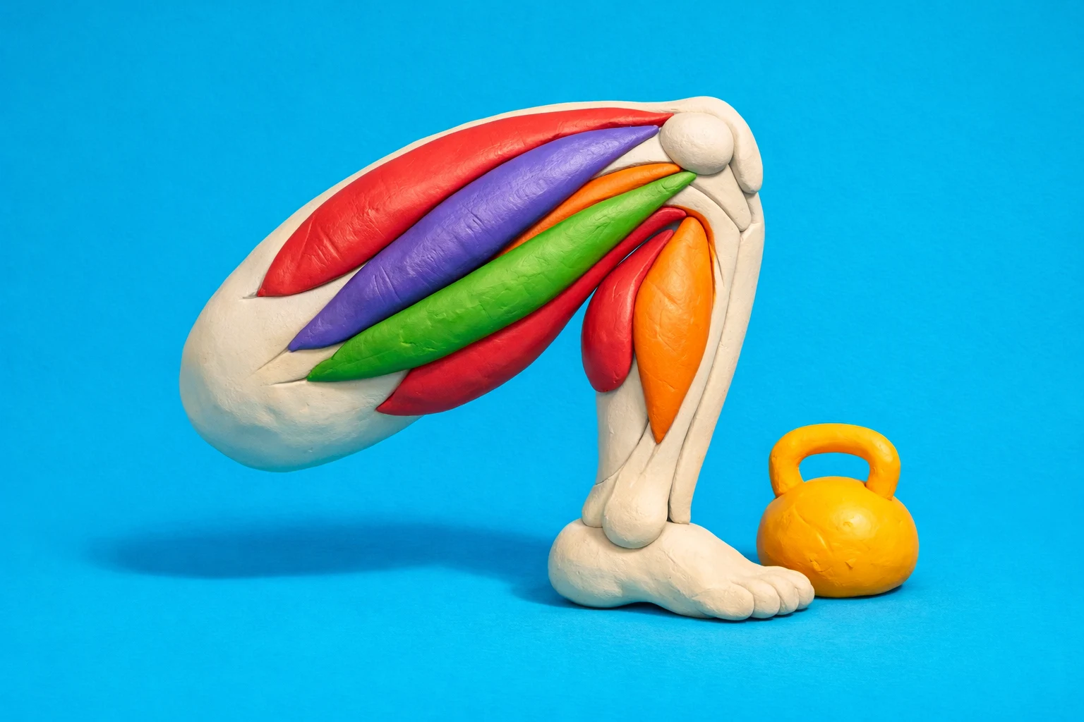
What is a good next practice step after I miss lower limb questions?
Add muscle attachment context and surface landmarks. Pair this quiz with Leg Muscles Practice Quiz for Lower Limb to connect bony landmarks like tibial tuberosity, fibular head, and greater trochanter to the structures that reference them.
Looking for more? Browse QuizWiz Education & Academics collection or explore the full compliance and training quizzes on QuizWiz.